For answers to common questions or to to learn more about your procedure, visit www.RadiologyInfo.org.
Interventional Services
Abscess drainage
- Non-surgical abdominal aortic aneurysm repair
- (AAA) Angiography Angioplasty & stent placement
- Cancer treatment – chemoembolization
- Chronic pelvic pain in women
- Deep Vein Thrombosis
- Gastrostomy (feeding tubes)
- Infertility Liver disease – cirrhosis
- Needle biopsy for diagnosis
- Peripheral vascular/arterial disease (PVD or PAD)
- Radiofrequency ablation for tumors
- Stroke
- Uterine Fibroid Embolization
- Vascular disease
- Venous disease

CT Exams
List of CT Exams available:
- Head
- IAC
- Temporal Bones
- Orbits Sinuses
- Facial Bones Neck, Chest
- Abdomen
- Pelvis
- Renal Stone CT Scan
- CT Arteriography for Pulmonary Embolus
- Extremities
- CT Aortography
- High-Resolution Lung CT
- Radiation Therapy Planning CT
Computed Tomography (CT)
Computed tomography (CT), pronounced tuh MAHG ruh fee, is an X-ray system used to produce images of various parts of the body, such as the head, chest, and abdomen. Doctors use CT images to help diagnose and treat diseases. The technique is also called computerized tomography or computerized axial tomography (CAT).
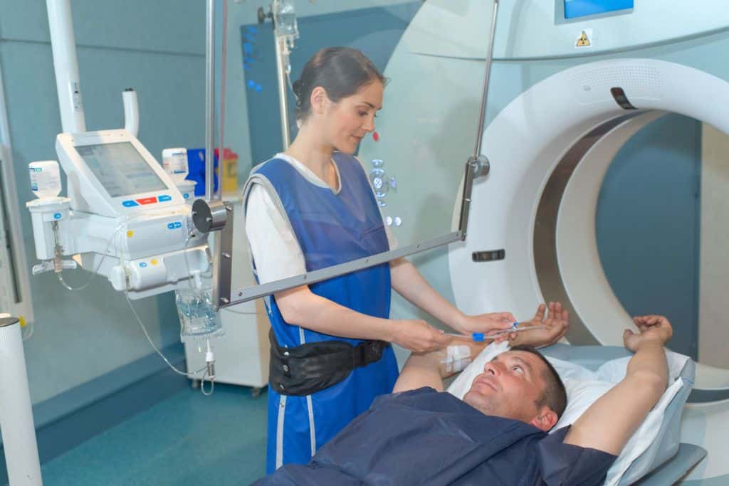
During a CT imaging procedure, a patient lies on a table that passes through a circular scanning machine called a gantry. The table is positioned so that the organ to be scanned lies in the center of the gantry. A tube on the gantry beams X-rays through the patient’s body and into special detectors that analyze the image produced. The gantry rotates around the patient to obtain many images from different angles. A computer then processes the information from the detectors to produce a cross-sectional image on a video screen. By moving the table in the gantry, doctors can obtain scans at different levels of the same organ. They can even put together several scans to create a three-dimensional computer image of the entire body.
Sometimes an iodine solution, called a contrast agent, is injected into the body to make certain organs or disease processes show up clearly in the CT scan. For example, the patient drinks a barium mixture to outline the inner surfaces of the stomach and bowel.
Doctors use CT scans to diagnose many conditions, such as tumors, infections, blood clots, and broken bones. CT also may be used to guide a biopsy needle into diseased tissue. In addition, it assists in treating some diseases that might otherwise require surgery. For example, doctors can use a CT scan to guide catheters (small tubes) to an abscess in the body and drain pus from the infected area.
Contributor: P. Andrea Lum, F.R.C.P.C., Abdominal Radiologist, Ottawa Civic Hospital, Ontario.
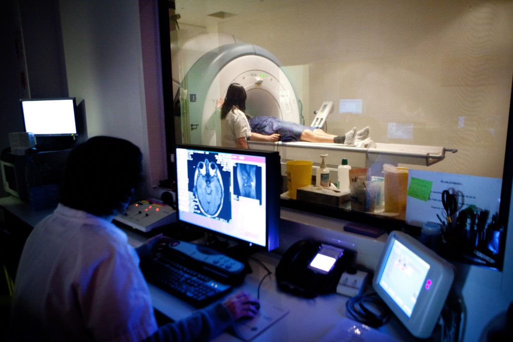
MRI Services
- Brain, orbits, head, and neck
- Cardiac MR
- Aortic MR
- Spine-cervical, thoracic, and lumbar
- Brachial Plexis
- Male and Female Pelvis
- Hips, Knees, ankles, and feet
- Soft tissue tumors
- TMJ MR
- MR shoulder orthography
- MR spectroscopy
- MR angiography of the brain, neck, chest, renal arteries, and lower extremities
- Breast masses and breast implants


Nuclear Medicine Imaging
- Bone, kidney, liver, spleen scan
- Hepatobiliary scan (gall bladder study)
- Nuclear lymphoscintiagraphy for sentinal node localization
- MUGA, Cardiac scans
- Radio-iodine therapy for thyroid cancer and Grave’s Disease
Routine Exams
- Esophagus
- Upper Gastrointestinal
- Barium Enema
- Intravenuous Pyleogram – IVP
- Conventional Radiography (X-rays)


Ultrasound Services
List of available Ultrasound Services:
- Carotids
- Doppler Thyroid Abdomen
- Gallbladder
- Liver
- Spleen Renal (Kidneys)
- Aorta Pelvis
- OB Testicular Extremities
Ultrasound
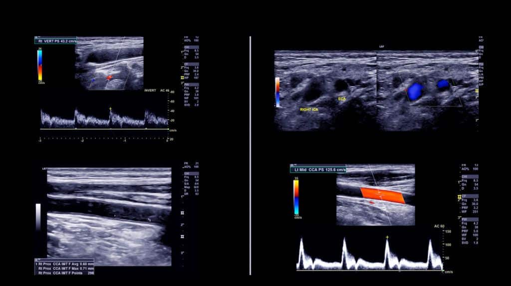
Complex devices called ultrasonic transducers to send ultrasound into liquids and solids, and receive echoes of the ultrasound. These devices convert electric energy into ultrasound and vice versa. Ultrasonic transducers are made of materials, such as quartz and some ceramics, that are piezoelectric (pronounced “pee ay zoh ih LEHK trihk”). This kind of material vibrates when an electric voltage is applied to it and produces a voltage when a sound wave causes it to vibrate.
Medical uses of ultrasonic transducers include machines that can detect the heartbeat of fetuses (unborn, developing babies). Other machines produce images of normal and diseased tissue inside the body, and images of fetuses. Physicians can use these machines to detect and evaluate cancer, fetal abnormalities, and other conditions. Instruments that use the Doppler effect can measure the flow of blood in the heart and blood vessels. A lithotripter uses pulses of ultrasound to break up gallstones or kidney stones.
Other uses of ultrasound include burglar alarms, automatic door openers, instruments that detect flaws in metal parts, machines that weld plastic, and tools that cut metal. Underwater sonar devices operate much like radar to measure distances to the ocean floor, detect submarines and other vessels, and even locate schools of fish. Ultrasonic cleaning instruments use sound waves to loosen dust and other contaminants from small, delicate products such as watches and electronic components.
Contributor: Frederick W. Kremkau, Ph.D., Professor and Director, Center for Medical Ultrasound, Wake Forest University School of Medicine.
Women's Services
- Breast Health
- Needle Localization
- Breast Ultrasound
- Mammogram
Women's Interventional Services
- fibroid embolization
- overian vein embolization for pelvic venous congestion
- Fallopian tube recanilization
- Non surgical embolization for post-operative bleeding


Vital 3D Imaging
Vital 3D Medical Imaging -Vital Images is medical imaging software that rapidly creates interactive three-dimensional images from two-dimensional images generated by CT and MR scanners, utilizing vital imaging (Vitrea) 3D workstation for
- 3D CT colonography
- 3D CT angiography
- 3D orthopedic bone evaluation
- 3D MR angiography3D cardiac angiography
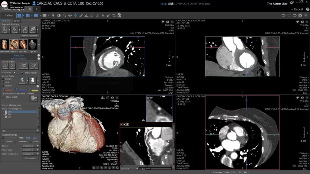
Christus St. Francis Cabrini hospital has recently purchased a Vital Images Vitrea 3D Imaging workstation Vitrea 2 is the name of the software which allows for fast and affordable computer 3D visualizations. Beginning with standard 2D CAT scan and magnetic resonance (MRI) images, Vitrea 2 allows radiologists and clinicians to quickly and easily analyze 3D views of human anatomy and most importantly non- invasively navigate in real-time through these images to better visualize and understand your internal anatomical structures. Ultimately, this software improves medical outcomes and reduces costs by helping physicians to diagnose conditions less invasively, plan surgical treatment with greater accuracy and detect certain cancers and cardiac diseases at an earlier stage. The radiologists of Central Louisiana Imaging utilize this advanced technology can improve the practice of medicine. Here are some examples of how this new technology will change how we practice medicine:
VIRTUAL COLONOSCOPY
This system allows virtual colonoscopy, real-time “fly-through” evaluation of the colon requiring only a colon cleansing prep, insufflation of the colon with air, followed by a CT scan. No I.V., sedation, day surgery, or recovery period is required. The entire procedure requires about 20-30 minutes to perform. Click HERE to experience a virtual colonoscopy fly-through.
3-D MR ANGIOGRAPHY
3-D CT ANGIOGRAPHY
Using a helical CT scanner, and a routine I.V. line for administration of I.V. contrast material, three-dimensional visualization of the vessels of the brain, neck, chest, and abdomen can be performed. This can be performed as an outpatient, without day surgery admission, and without any groin arterial puncture or recovery time. Click HERE to see a 3-D angiogram of the heart and aorta.
3-D CARDIAC ANGIOGRAPHY
3-D ORTHOPEDIC BONE EVALUATION
The three-dimensional analysis of complicated fractures, dislocations, or other orthopedic problems can be crucial to the Orthopedic surgeon in the evaluation and pre-surgical planning of complicated orthopedic injuries. Click HERE to see an example study evaluating the position of an orthopedic plate relative to the bones and tendons of the foot.

